
Dog skeleton 101 Dog Anatomy Bones Animal Hackers
Skeleton of the dog. Axial Skeleton. = the Head and spinal column. Head - made up of around 50 bones. Skull - occipital crest along top, occiput - rear point of skull zygomatic arch - under and around the base of the eye. Muzzle - nasal crest (largely cartilaginous) top of nose. - maxilla - top jaw. mandible - bottom jaw.

Anatomy Of Dog Spine Canis Lupus German Shepherd Skeleton Dog sketch, Dog anatomy, Dog skeleton
2021 Ultimate Guide to Dog Anatomy. As the pace of veterinary advancement accelerates, even the most experienced veterinary teams are challenged to keep up with all the changes that impact their practice.. potential outcomes and prognoses and tools e.g. anatomical diagrams, clinical formulas and programs and home care videos; Enhancing the.

Dog skeleton with major bone elements labeled (Davis, 1987, p. 54;... Download Scientific Diagram
Whereas giant breeds can take between 18 months and 2 years for their growth plates to fuse. Speaking of skeletons, a dog has 320 bones in their body (depending on the length of their tail) and around 700 muscles. Muscles attach to bones via tendons. Depending on the breed of dog, they will have different types of muscle fibers.
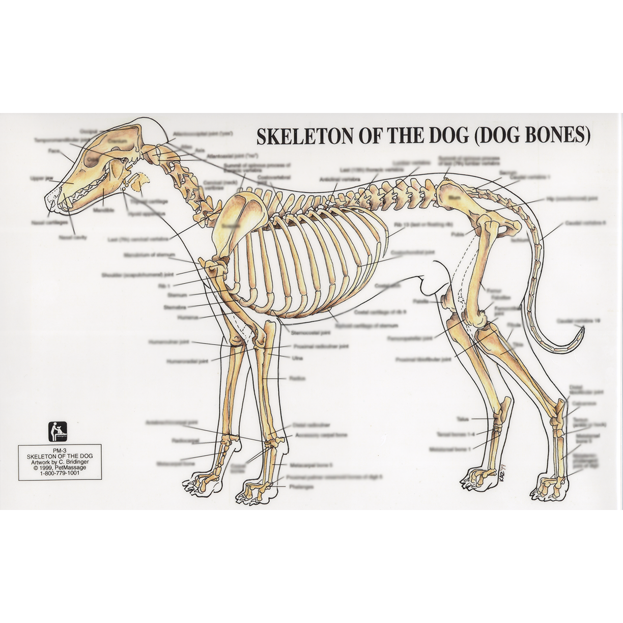
PetMassage™ Chart 3 Skeleton of the Dog · PetMassage™ Training and Research Institute
Below is a diagram of a dogs anatomy: The coat of a dog varies in colors ranging from all black, brown, beige and white to others being of a mix with light or dark markings and colorations on different parts of their bodies and faces. Their fur ranges from short, smooth fur to long shaggy fur to soft and fluffy to hard and coarse.
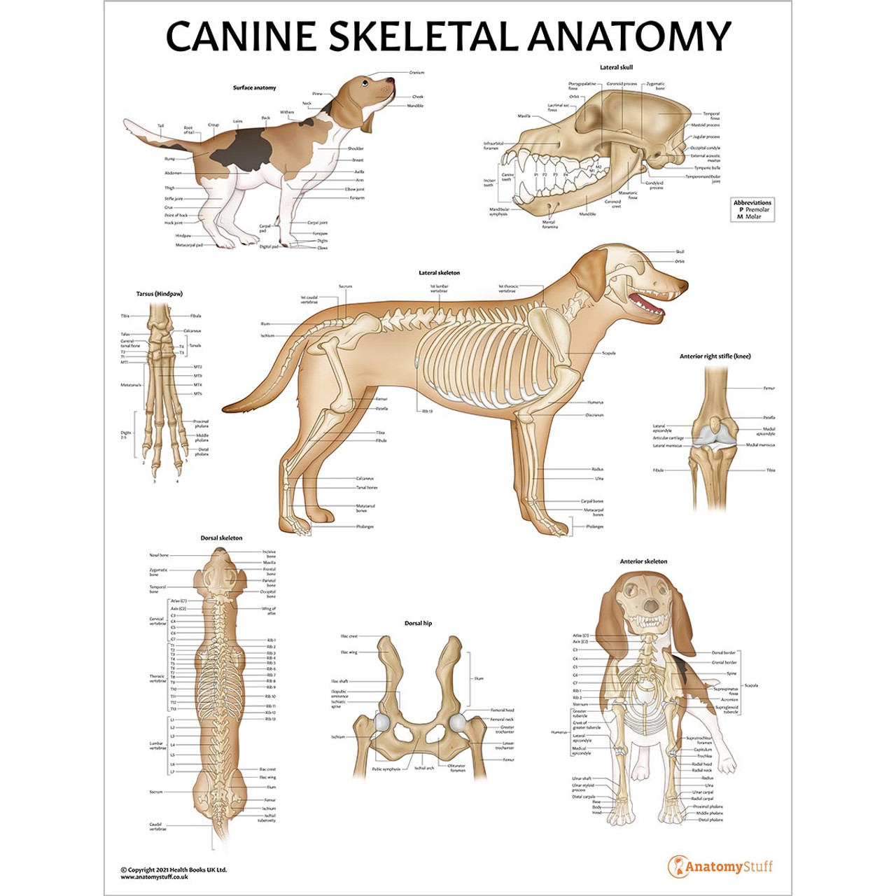
Canine Skeletal Anatomy Laminated Chart Dog Skeleton Poster
Anatomy atlas of the canine general anatomy: fully labeled illustrations and diagrams of the dog (skeleton, bones, muscles, joints, viscera, respiratory system, cardiovascular system). Positional and directional terms, general terminology and anatomical orientation are also illustrated.

Dog skeleton with major bone elements labeled (Davis, 1987, p. 54;... Download Scientific Diagram
In addition, in the diagram, you will find a few identified skull bones. The sternum and the ribs are also identified in the dog skeleton labeled diagram. If you want to more updated dog skeleton labeled diagram, you may join anatomy learner on social media (get more images). Frequently asked questions on dog skeleton bones.
Dog Skeletal Anatomy
ISSN 2534-5087. This veterinary anatomy module of the dog contains 218 illustrations dedicated to the canine osteology anatomy. Here are presented scientific illustrations of the canine skeleton, with the main dog's bones and its structures displayed from different anatomical standard views (cranial, caudal, lateral, medial, dorsal, palmar..).

Labeled atlas of anatomy illustrations of the dog Bones Skeletal system Dog skeleton
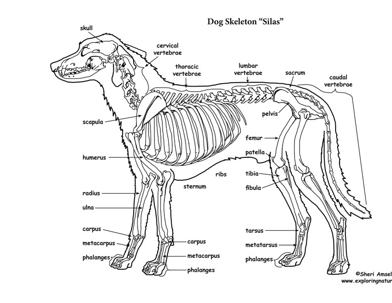
The skeleton is composed of the hard tissues of the body, and its primary functions are to support the body, to provide a system of levers used in locomotion, to protect the soft organs of the body, and to produce red blood cells (hematopoiesis). A dog's skeleton is formed so the dog can run fast, hunt and chase.

Dog Anatomy Dog Skelton
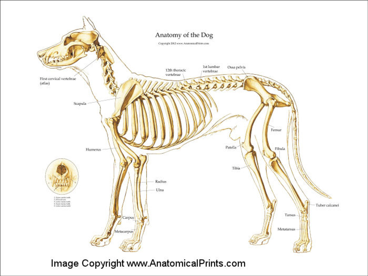
Animal Anatomy (Veterinary Diagrams) Dog Skeletal Anatomy. High Resolution PDF for Printing. Click Here. Link to More Information About This Animal. Click Here. Citing Research References. When you research information you must cite the reference. Citing for websites is different from citing from books, magazines and periodicals.

Labeled atlas of anatomy illustrations of the dog Bones Skeletal system Molecular Shapes
It provides information about a dog's skeletal, reproductive, internal, and external anatomy, along with accompanying labeled diagrams. After mating, dogs experience something called a copulatory tie, wherein they remain in the coital position. The male dog dismounts the female at this time. The dogs can remain in this position from a few.

Vintage 1935 Dog Veterinary Print Skeleton Of Dog Anatomy Of Dog Canine Skeleton Dog Bones Book
Anatomic Planes. The main planes of motion for dogs are as follows (see Figure 5-1): • The sagittal plane divides the dog into right and left portions. If this plane were in the midline of the body, this is the median plane or median sagittal plane. • The dorsal plane divides the dog into ventral and dorsal portions.
Canine Skeleton Poster Clinical Charts and Supplies
Dog skeleton. As with any vertebrate animal, the skeleton of a dog has the function of supporting the body for movement and protecting its internal organs. We can divide the canine skeleton into three main sections: Axial skeleton: skull, spine, ribs and sternum bones. Appendicular skeleton: bones of the extremities.

Dog Skeleton Anatomy by TheDragonofDoom on DeviantArt
This module of vet-Anatomy presents an atlas of the anatomy of the head of the dog on a CT. Images are available in 3 different planes (transverse, sagittal and dorsal), with two kind of contrast (bone and soft tissues).
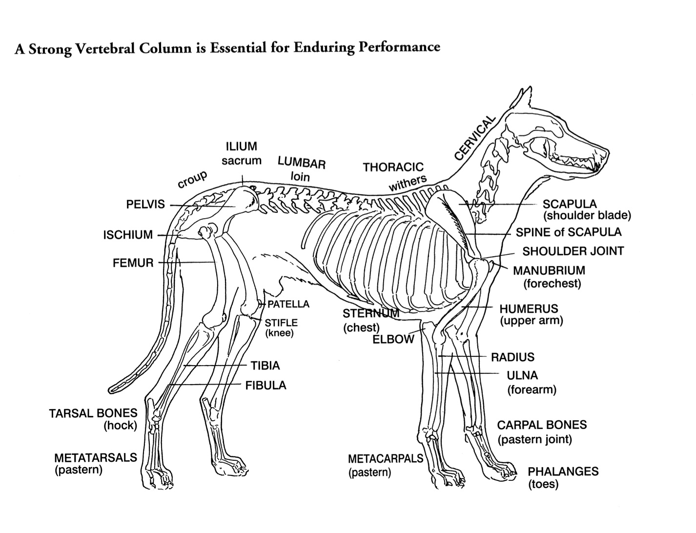
Helen King on Structure Evaluation Susan Garrett's Dog Training Blog
Dog anatomy comprises the anatomical studies of the visible parts of the body of a domestic dog.Details of structures vary tremendously from breed to breed, more than in any other animal species, wild or domesticated, as dogs are highly variable in height and weight. The smallest known adult dog was a Yorkshire Terrier that stood only 6.3 cm (2.5 in) at the shoulder, 9.5 cm (3.7 in) in length.

FileDog anatomy lateral skeleton view.jpg
Canine Anatomy The Dog. Dogs are domesticated descendants of the wolf and belong to the canine species. Dogs are loyal, lovable animals that make ideal companions. Their physical characteristics and temperaments vary from breed to breed, but they all share anatomical characteristics and exceptional, highly developed senses. Canine Skeleton

Anatomy of a male dog crosssection, showing the skeleton and internal organs. Colour process
Components of the Musculoskeletal System in Dogs. Bones provide rigid structure to the body and shield internal organs from damage. They also house bone marrow, where blood cells are formed, and they maintain the body's reservoirs of calcium and phosphorus. Old bone tissue is constantly replaced with new bone tissue in a process called.
